A promising approach to intraoperative cancer diagnosis

The figures depict images of invasive ductal carcinoma, mucinous carcinoma, and papillary carcinoma, respectively. Despite the variations in tissue structures among different tumor types, the activated regions of the model corresponded to the areas of yellow malignant cell aggregation. Scale bar = 100 µm. Credit: Science China Press Rapid and accurate intraoperative diagnosis is critical for…

Read More

Newly identified PET biomarker predicts success of immune checkpoint blockade therapy

PET imaging of 124I-αGal-1 predicts ICB therapy. Credit: N Liu and X Yang, et al. Peking University, Beijing, China. The protein galectin-1 (Gal-1) has been identified as a new PET imaging biomarker for immune checkpoint blockade (ICB) therapy, allowing physicians to predict the tumor responses before beginning treatment. Information garnered from Gal-1 PET imaging could…

Read More

Ultrasound can help patients with a type of rheumatic disease lead longer and healthier lives

Credit: MART PRODUCTION from Pexels A dissertation at Umeå University shows that ultrasound can help patients with a type of rheumatic disease to live longer and healthier lives. These patients have so far had an increased risk of cardiovascular disease, which contributes to premature death. Patients with a type of rheumatic disease called radiographic axial…

Read More

Deep learning-based whole-body PSMA PET/CT attenuation correction utilizing Pix-2-Pix GAN

AI-generated PET results shown overlaid on CT. Credit: Oncotarget (2024). DOI: 10.18632/oncotarget.28583 A new research paper was published in Oncotarget, titled “Deep learning-based whole-body PSMA PET/CT attenuation correction utilizing Pix-2-Pix GAN.” Radiation dosage limits the sequential PET/CT studies oncology patients can undergo during their treatment follow-up course. In this new study, researchers from the National…

Read More

Low-cost MRI paired with AI produces high-quality results

First introduced five decades ago, MRI scanners are now a cornerstone of modern medicine, vital for diagnosing strokes, tumors, spinal conditions and more, without exposing patients to radiation. A magnetic resonance imaging device built with off-the-shelf parts and paired with AI matched the performance of high-end MRI machines, according to a study published Thursday that could…

Read More

Study introduces hyperspectral dark-field microscopy for rapid and accurate identification of cancerous tissues

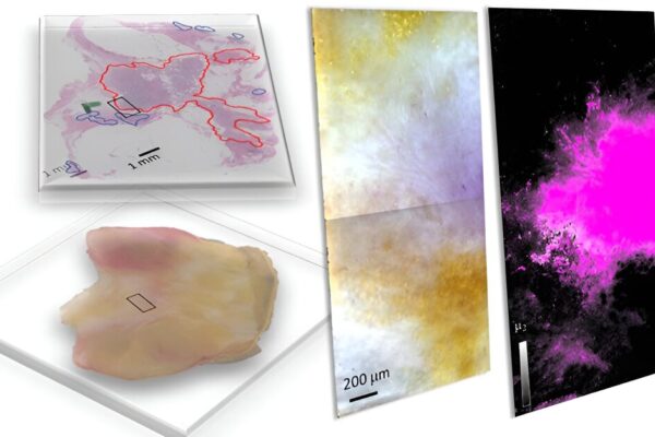
Freshly dissected tissue (lower left) and its pathology-prepared slide with identified tumor regions by a pathologist (upper left), and a pseudo-color image of hyperspectral dark-field microscopy (HSDFM) data cube (middle) region marked on the tissue images on the left. The HSDFM generates an image where each pixel contains a spectral signature corresponding to tissue composition….

Read More